List of projects of the team
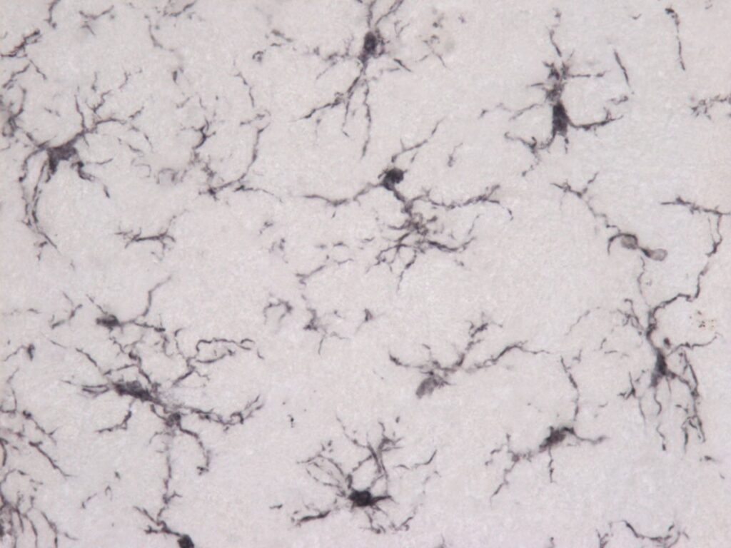
Segmenting microglial cells from microscopical images for morphological analysis

Microglia are the resident immune cells of the central nervous system, taking up around 10% of all brain cells. In most cases, they are the first to react to injury, inflammation, or any neurodegenerative issues, and, according to traditional theory, can show either neurotoxic or neuroprotective behavior. Changes in function mean changes in morphology in the case of microglia: they can undergo remarkable phenotypical changes with unmatched dynamics. Understanding these dynamics can take us closer to understanding diseases with altered microglial behavior, such as Alzheimer’s disease or amyotrophic lateral sclerosis.
The project focuses on providing accurate segmentation of glial cells from microscopic images, offering reliable and detailed image-derived data for further analysis of the area of interest.
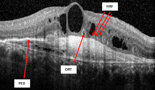
Enumerating biomarkers on oct Images for optimizing AMD treatment

Age-related Macular Degeneration (AMD) is one of the leading causes of vision loss in the Western world. It mostly affects elderly people causing a loss of sight.
The project focuses on characterizing biomarkers of AMD in Optical Coherence Tomography (OCT) images to analyze the evolution of the disease and help develop methods for better treatment and early detection.
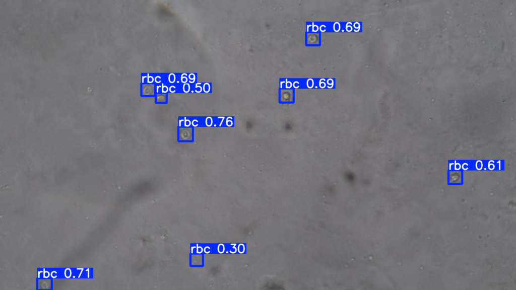
Analysis of Urine sediment microscopy images

Reaching a firm diagnosis of a nephrology-related disease is a hard process, that oftentimes involve examination of the microscopy images of urine sediment.
The project aims to speed up and enhance the analysis process by extracting useful information from microscopy images.